TSH, Free T4, and Free T3: The Cellular Science Behind Your Thyroid Panels
When a physician orders a thyroid panel, the resulting printout typically lists a series of abbreviations: TSH, Free T4, and Free T3. To the average reader, these are merely numbers on a page with a reference range next to them. However, inside your body, these values reflect a highly coordinated, microscopic dance of molecular synthesis, transport, and negative feedback.
To truly understand your thyroid health, we must peer past the lab slip and look directly at the cellular machinery of the thyroid gland itself.
The Master Controller: What TSH Actually Measures
Your thyroid gland does not operate in isolation. It is governed by a small, bean-sized gland at the base of your brain: the anterior pituitary gland. When your brain senses that circulating thyroid hormone levels are dropping, the hypothalamus releases Thyrotropin-Releasing Hormone (TRH). This tripeptide signals the pituitary to secrete Thyroid-Stimulating Hormone (TSH), also known as thyrotropin.
At the biochemical level, TSH is a complex glycoprotein composed of two non-covalently linked peptide subunits:
- The Alpha (α) Subunit: This subunit contains an identical amino acid sequence to other pituitary glycoprotein hormones, including Follicle-Stimulating Hormone (FSH) and Luteinizing Hormone (LH).
- The Beta (β) Subunit: This subunit is unique to TSH and confers its specific physiological action, allowing it to bind exclusively to TSH receptors on thyroid follicular cells.
When TSH binds to these receptors, it acts as a systemic “gain control”. It does not simply turn the thyroid “on” or “off”; rather, it amplifies or dampens the rate of chemical reactions across almost every cell in your body, setting your baseline metabolic rate.
Your thyroid gland does not operate in isolation. It is governed by a small, bean-sized gland at the base of your brain: the anterior pituitary gland. When your brain senses that circulating thyroid hormone levels are dropping, the hypothalamus releases Thyrotropin-Releasing Hormone (TRH). This tripeptide signals the pituitary to secrete Thyroid-Stimulating Hormone (TSH), also known as thyrotropin.
At the biochemical level, TSH is a complex glycoprotein composed of two non-covalently linked peptide subunits:
- The Alpha (α) Subunit: This subunit contains an identical amino acid sequence to other pituitary glycoprotein hormones, including Follicle-Stimulating Hormone (FSH) and Luteinizing Hormone (LH).
- The Beta (β) Subunit: This subunit is unique to TSH and confers its specific physiological action, allowing it to bind exclusively to TSH receptors on thyroid follicular cells.
When TSH binds to these receptors, it acts as a systemic “gain control”. It does not simply turn the thyroid “on” or “off”; rather, it amplifies or dampens the rate of chemical reactions across almost every cell in your body, setting your baseline metabolic rate.
Inside the Thyroid Follicle: How Hormone is Made
The functional units of your thyroid are microscopic, hollow vesicles called thyroid follicles. These follicles are lined by a single layer of simple cuboidal epithelial cells and are filled with a sticky, protein-rich fluid called thyroid colloid
To synthesize thyroid hormones, your follicular cells must perform a series of highly specialized steps:
1. Iodine Trapping and the NIS Symporter
Thyroid hormones are entirely unique in biology because they require large quantities of the trace element iodine to function. Your thyroid traps iodide against a steep concentration gradient (often making cellular iodide 25 to 50 times more concentrated than blood plasma). This is achieved by an energy-dependent pump in the basolateral membrane called the Sodium/Iodide Symporter (NIS). The NIS couples the inward transfer of two sodium ions down their favorable electrochemical gradient with one iodide ion. This sodium gradient is actively maintained by the continuous expenditure of ATP via the sodium/potassium ATPase pump.
Certain competing anions, such as perchlorate, pertechnetate, and thiocyanate, can bind to the NIS and block iodide uptake, a property clinicians exploit for diagnostic and therapeutic purposes.
2. Apical Transport and Thyroglobulin Synthesis
Once inside the follicular cell, iodide must cross to the apical membrane to enter the colloid lumen. It does so via a sodium-independent iodide transporter called pendrin. Simultaneously, the rough endoplasmic reticulum of the follicular cell synthesizes a massive dimeric glycoprotein called thyroglobulin (Tg) (molecular mass of 660,000 Da). Thyroglobulin is packaged into secretory vesicles and discharged across the apical membrane into the follicle’s central lumen.
3. Organification and Coupling via TPO
Once inside the colloid, iodide must be oxidized to react with the tyrosine residues in thyroglobulin. This critical step is catalyzed by the apical membrane-bound enzyme thyroid peroxidase (TPO) in the presence of hydrogen peroxide (H2O2). The H2O2 is generated by a calcium-dependent NADPH oxidase present in the apical brush border.
TPO performs two major functions:
- Organification: It attaches iodine molecules to the tyrosine residues of thyroglobulin, forming monoiodotyrosine (MIT) and diiodotyrosine (DIT).
- Coupling: It couples these iodinated tyrosines together while they are still in peptide linkage within the thyroglobulin molecule. Coupling two DIT molecules yields thyroxine (T4). Coupling one MIT and one DIT yields triiodothyronine (T3).
Free vs. Bound: The Transport Dilemma
Once synthesized, thyroid hormones are stored extracellularly in the follicular colloid as part of the thyroglobulin molecule. Under TSH stimulation, follicular cells engulf colloid via endocytosis, fuse these colloid droplets with lysosomes, and proteolytically digest thyroglobulin to release free T4 and T3 into the blood.
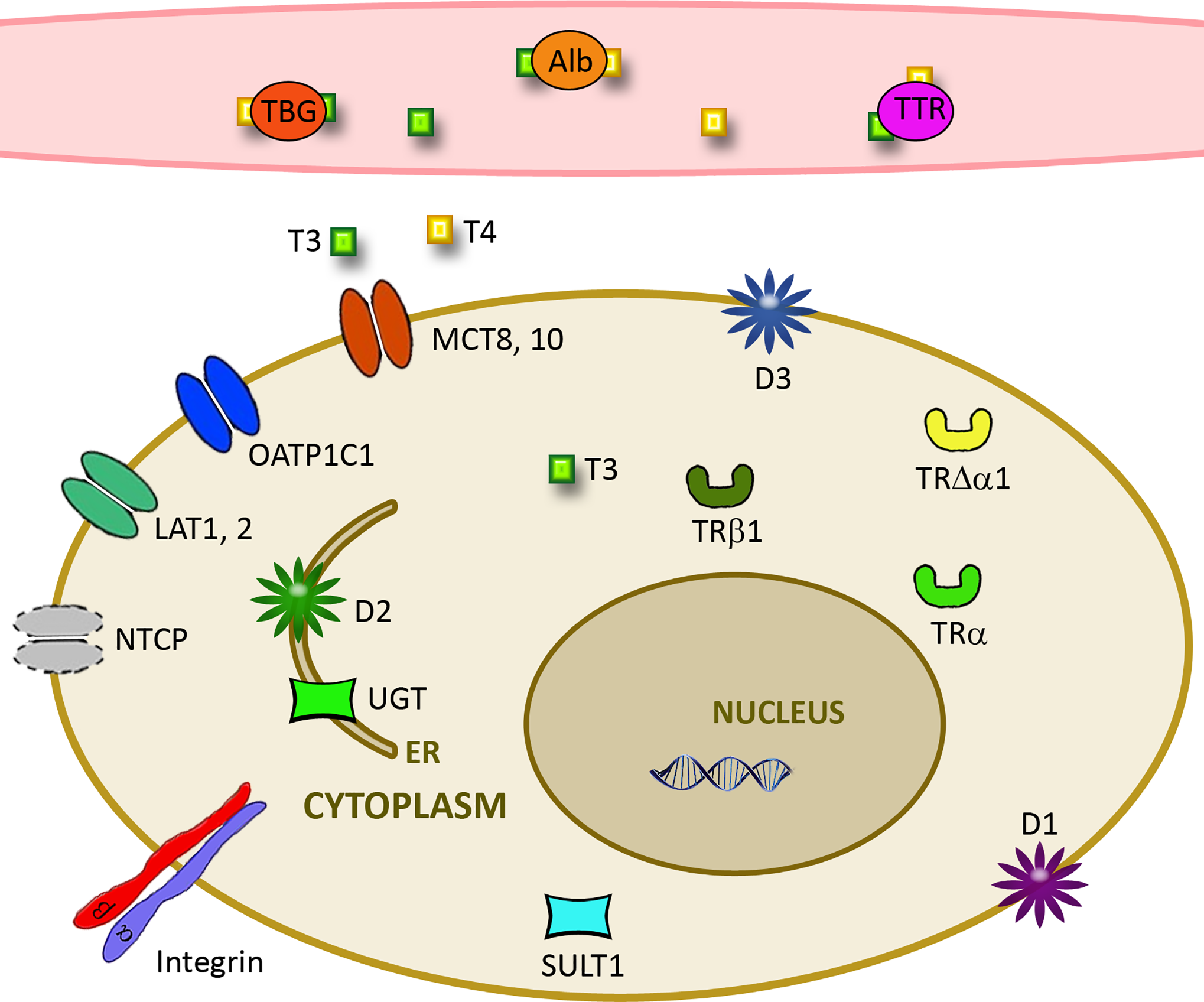
However, thyroid hormones are highly hydrophobic (water-insoluble) and cannot travel freely through the bloodstream in large amounts. Therefore, more than 99% of circulating T4 and T3 is bound to carrier proteins synthesized by the liver, principally Thyroxine-Binding Globulin (TBG), transthyretin (TTR), and albumin.
This protein-bound pool acts as an inactive reservoir that shields the hormone from rapid degradation, maintaining a stable supply. Only the unbound, or “Free” portion (<1%) can cross cell membranes to bind to nuclear receptors and produce biological activity.
If your liver produces more TBG (such as during pregnancy due to elevated estrogens), the total T4 in your blood will rise, but your Free T4 will remain tightly regulated and normal. This is why measuring Free T4 and Free T3 is clinically superior to measuring Total T4 and Total T3.
Why T4 is a Prohormone and T3 is the Active Driver
Your thyroid gland secretes T4 and T3 in a ratio of approximately 20:1. However, T4 is largely considered a prohormone. While it is highly stable in blood (with a half-life of about 6.2 days), its affinity for nuclear thyroid receptors is exceptionally low.
To exert its metabolic effects, T4 must be converted to T3 (half-life of 1.0 day). This transformation occurs in peripheral tissues—especially the liver and kidneys—and is catalyzed by selenium-containing enzymes called deiodinases, which strip a single iodine atom from the outer ring of T4. Approximately 85% of the active T3 circulating in your body is generated by this peripheral conversion rather than direct secretion from the thyroid gland.
How to Interpret Thyroid Blood Tests Together
Because the hypothalamus, pituitary, and thyroid operate on a tightly calibrated negative feedback loop, a single lab number rarely tells the whole story. The pituitary constantly monitors circulating levels of free T4 and T3. If levels rise, the pituitary suppresses TSH; if levels fall, TSH rises exponentially.
| Clinical Scenario | TSH Level | Free T4 Level | Free T3 Level | What is Happening at the Cellular Level? |
|---|---|---|---|---|
| Primary Hypothyroidism | High | Low | Low or Normal | Glandular failure (often autoimmune destruction). The pituitary is frantically sending TSH signals, but the thyroid follicles are too damaged to synthesize hormones. |
| Primary Hyperthyroidism | Low | High | High | Glandular overactivity. Excess free hormone circulating in the blood binds to pituitary thyrotropes, completely shutting down TSH production. |
| Subclinical Hypothyroidism | High | Normal | Normal | Early-stage thyroid struggle. Circulating free hormones are still within normal limits, but the pituitary must work harder (producing more TSH) to maintain that baseline. |
| Central Hypothyroidism | Low or Normal | Low | Low | Upstream failure. The thyroid gland is fully capable of producing hormone, but the pituitary or hypothalamus is damaged (e.g., by a tumor) and fails to send the TSH signal. |
However, the human endocrine system is highly individualized, and thyroid hormone levels can fluctuate based on stress, concurrent illness, medication timing, and nutritional status. A single set of blood test numbers should never be used to self-diagnose or self-treat. One always needs to interpret lab work in consultation with a certified endocrinologist or who can correlate these values with your clinical symptoms.
References:
Pellegrini, S., & Pietrini, P. (2023). Clinical biochemistry of the mind. In M. Ciaccio (Ed.), Clinical Biochemistry and Laboratory Medicine (pp. 553–564). Springer. https://doi.org/10.1007/978-3-031-24958-7_41
Fröhlich, E., & Wahl, R. (2021). Physiological role and use of thyroid hormone metabolites – potential utility in COVID-19 patients. Frontiers in Endocrinology, 12. https://doi.org/10.3389/fendo.2021.587518