A Simple Guide and Memory Trick
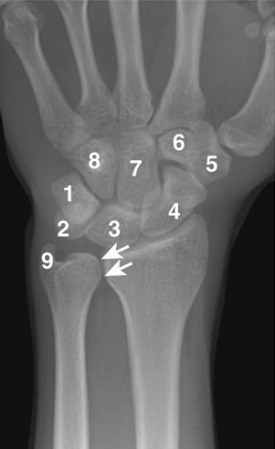 | 1, triquetral; 2, pisiform; 3, lunate; 4, scaphoid; 5, trapezium; 6, trapezoid; 7, capitate; 8, hamate; 9, ulnar styloid; DRUJ stands for distal radioulnar joint – the joint between the radius and ulna near the wrist. |
Figure 1 AP radiograph of the wrist as presented in Waldman and Campbell (2010)
The wrist is made up of eight small carpal bones arranged in two rows. Learning their names and positions is an important part of anatomy, particularly for students studying medicine, physiotherapy, nursing, sports science, and other healthcare subjects.
The easiest way to learn the carpal bones is to understand their arrangement and use a mnemonic to help recall them.
1. What Are the Carpal Bones?
The eight carpal bones connect the forearm to the hand. They sit between the distal ends of the radius and ulna and the bases of the five metacarpal bones. The carpal bones are arranged into a proximal row and a distal row.
When looking at the hand with the palm facing you, the bones can be remembered from thumb side (radial) to little-finger side (ulnar).
Proximal Row
From radial to ulnar:
- Scaphoid
- Lunate
- Triquetrum
- Pisiform
- Distal Row
From radial to ulnar:
- Trapezium
- Trapezoid
- Capitate
- Hamate
A useful way to visualise the arrangement is:
Proximal: Scaphoid → Lunate → Triquetrum → Pisiform
Distal: Trapezium → Trapezoid → Capitate → Hamate
2. The Classic Carpal Bones Mnemonic
A popular mnemonic for remembering the eight bones is:
Some Lovers Try Positions That They Can’t Handle
The first letter of each word corresponds to a carpal bone:
| Mnemonic | Carpal bone |
| Some | Scaphoid |
| Lovers | Lunate |
| Try | Triquetrum |
| Positions | Pisiform |
| That | Trapezium |
| They | Trapezoid |
| Can’t | Capitate |
| Handle | Hamate |
This mnemonic follows the bones from radial to ulnar, first through the proximal row and then through the distal row.
3. How to Tell the Bones Apart
Knowing the names is useful, but recognising a few characteristic features can make the anatomy much easier to remember.
1. Scaphoid
The scaphoid is the most lateral bone of the proximal row, on the thumb side. Its name is associated with a boat-like shape. It also bridges the proximal and distal carpal rows.
Clinically, the scaphoid is particularly important because it is the most commonly fractured carpal bone. A fall on an outstretched hand can transmit force through the scaphoid, and fractures may cause pain and tenderness around the anatomical snuffbox.
The scaphoid also has a vulnerable blood supply. Following certain fractures, particularly through its waist, the proximal part can be at risk of avascular necrosis because of its limited blood supply.
Memory tip:
Scaphoid = Snuffbox + Scaphoid fracture
2. Lunate
The lunate sits immediately medial to the scaphoid and has a crescent-like shape.
Its position is clinically significant because it lies close to the median nerve. A lunate dislocation can therefore contribute to compression of the median nerve within the carpal tunnel.
Memory tip:
Lunate = Luna = Moon = crescent shape
3. Triquetrum
The triquetrum is the third bone in the proximal row. It has a roughly pyramid-shaped configuration and articulates with the lunate, pisiform, and hamate.
Memory tip:
Think “Tri” when remembering its position as the third bone in the proximal sequence.
4. Pisiform
The pisiform is the small, pea-shaped bone at the ulnar side of the proximal row. It articulates with the triquetrum and provides an attachment site for tendons and ligaments.
Memory tip:
Pisiform = pea-shaped
The Distal Row
5. Trapezium
The trapezium is the most lateral bone in the distal row and is located beneath the base of the thumb.
One of its most important features is its saddle-shaped articulation with the first metacarpal. This joint plays an important role in thumb movement and therefore contributes to the thumb’s ability to oppose the fingers.
Memory tip:
Trapezium = Thumb
6. Trapezoid
The trapezoid lies between the trapezium and capitate. It is the smallest bone in the distal carpal row.
Memory tip:
Remember that trapezium and trapezoid are neighbours.
7. Capitate
The capitate is the largest and most centrally located carpal bone. It articulates with several surrounding bones and provides attachment points for intercarpal ligaments.
Memory tip:
Capitate = central + largest
8. Hamate
The hamate forms the ulnar border of the distal carpal row. It has a distinctive hook-like projection, called the hamulus or hook of the hamate.
The hook is clinically important because it is involved in the anatomy around Guyon’s canal, where the ulnar nerve and artery pass.
Memory tip:
Hamate = Hook
A Quick Way to Learn All Eight
Instead of trying to memorise eight unrelated names, break them into two groups:
Proximal row:
S L T P
Distal row:
T T C H
Then attach the mnemonic:
Some Lovers Try Positions | That They Can’t Handle
If you can remember SLTP | TTCH, you can reconstruct the names of all eight carpal bones.
4. Why the Carpal Bones Matter Clinically
The carpal bones are not simply a collection of small bones. Their arrangement creates a complex wrist structure that allows the hand to move while providing stability.
The carpal arrangement contributes to the carpal tunnel, with the carpal bones forming its floor and supporting the flexor tendons and median nerve. The scaphoid and trapezium have prominent tubercles that contribute to the anterior boundaries of the tunnel.
Several carpal bones also have important clinical associations:
Scaphoid: commonly fractured and vulnerable to avascular necrosis.
Lunate: dislocation can affect the nearby median nerve.
Hamate: fractures of the hook can cause ulnar-sided symptoms.
Carpal instability: disruption of the relationships between the bones and their ligaments can produce patterns such as DISI and VISI.
5. Final Memory Trick
When revising for an anatomy exam, remember:
“Some Lovers Try Positions That They Can’t Handle.”
Then visualise the two rows:
Scaphoid – Lunate – Triquetrum – Pisiform
Trapezium – Trapezoid – Capitate – Hamate
Or simply:
SLTP | TTCH
Once you know the sequence, add the distinctive features:
Scaphoid = snuffbox
Lunate = moon-shaped
Triquetrum = three
Pisiform = pea-shaped
Trapezium = thumb
Trapezoid = small neighbour
Capitate = largest/central
Hamate = hook
This combination of sequence + visual features + clinical associations makes the eight carpal bones much easier to recall during exams and practical anatomy assessments.
References
This article is based on Tang A, Varacallo MA, “Anatomy, Shoulder and Upper Limb, Hand Carpal Bones,” StatPearls, NCBI Bookshelf.
Tang, A., & Varacallo, M. (2020). Anatomy, Shoulder and Upper Limb, Hand Carpal Bones. In PubMed. StatPearls Publishing. https://www.ncbi.nlm.nih.gov/books/NBK535382/
Waldman, S. D, & Campbell, R. S. D. (2010). Imaging of Pain. Saunders.